About The Xerra
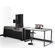
The Xerra is a next generation cryo-fluorescence tomography tool that allows for comprehensive 3D fluorescence & anatomical images from 2D images. The Xerra does not require any radio-labeling and is located only inside the ARCF facility.
Features and Benefits
Superior resolution down to 20 um and up to 100 um in sections from 10 um to 50 um in thickness
High sensitivity to detect signals deep in tissue with an optical field of view of maximum 24 x 14 cm and minimum of 8 x 5 cm
High throughput, with the capacity to process several mice or multiple dissected organs on the same block
Reduces data variability by combining CFT with in vivo imaging and tissue microscopy from the same animal for reconstruction of whole body/tissue data in white and fluorescent light images
Streamlined user process, with no fixation, perfusion, tissue clearing, or radio-labeling required
Ability to select specific slices and transfer onto histology tape for further analysis
Maintains complete anatomical context with a single machine
Allows for versatile visualization from 470-780 nm
Automated workflows that obtain automated reconstructions of 2D slices into 3D as well as map exact anatomical location of molecular imaging data
Has integrated software and an onboard computer that allows for fast quantitative analysis, import/export function and provides access to open source data that can be integrated with iPACS
Applications
Imaging/characterization of tumor environment
Drug characterization
ADME information and visualization of whole animal
High resolution visualization of cell clusters, isolated organs and whole animals
Visualization of vector distribution & vector mediated gene expression
If you are interested in using the Xerra, please contact Darshini for more information about pricing.
Contact
Darshini Vijayakumar
Research Scientist I | University of Washington
Department of Radiology | Imaging Research Laboratory
Lab Manager | Radiology Optical Imaging Core
Portage Bay Building, Room 222
Seattle, WA 98101
dvijay26@uw.edu